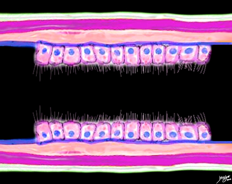
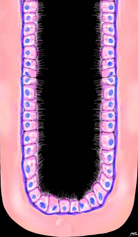
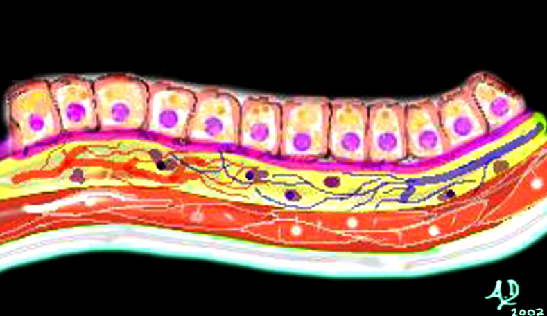
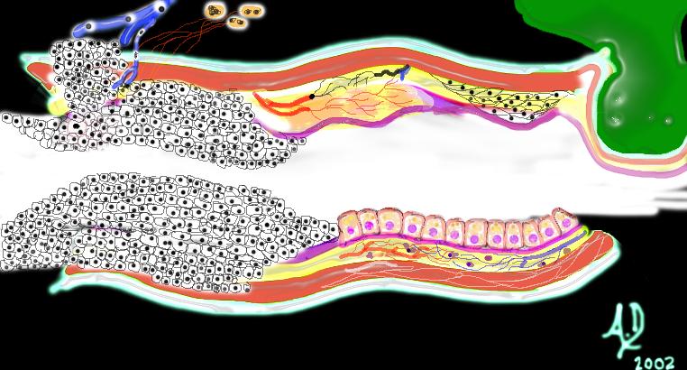
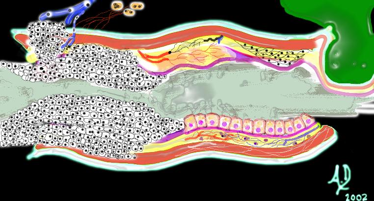
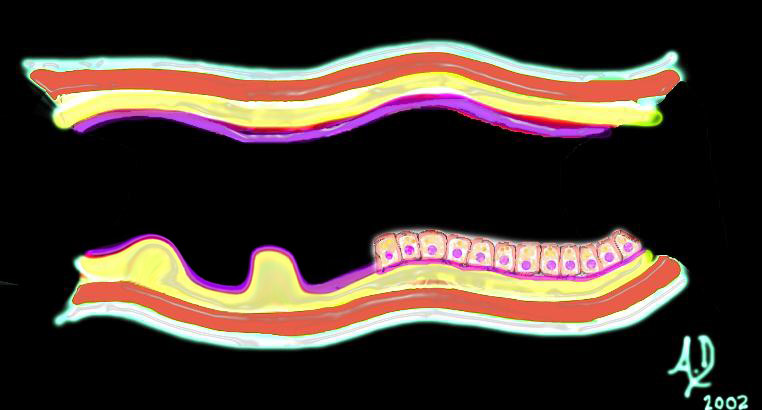
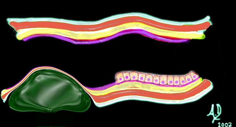
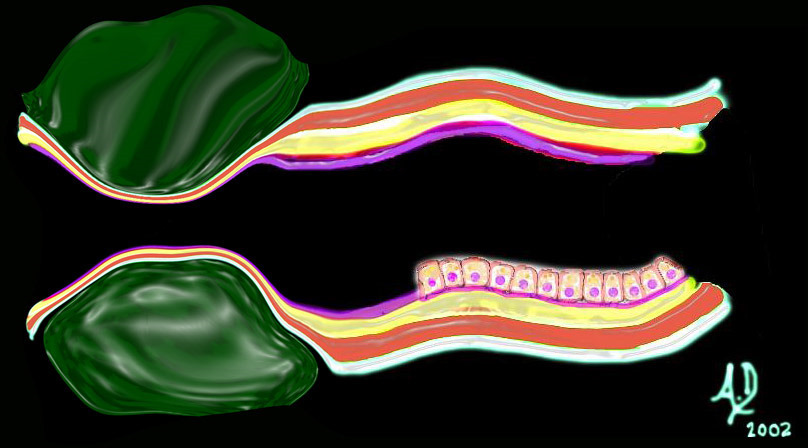
The diagram typifies the histological appearance of the endometrium which follows the basic 3 component pattern of tubular systems in the body; mucosa – ciliated columnar cells, basement membrane (blue) and submucosa (orange) muscularis – (pink) and serosa (cream). code uterus the common vein endometrial cavity endometrial stripe epithelium histology ciliated columnar epithelium basement membrane submucosa muscularis muscular layer histology cytologyThe higher power diagram reveals the cytological appearance of the simple tubular gland of the endometrial lining of the uterus showing the ciliated columnar cells resting on the basement membrane (deep blue) which in turn are embedded in a stroma (pink) code uterus the common vein endometrial cavity endometrial stripe epithelium histology ciliated columnar epithelium basement membrane histology cytology