The Secondary Lobule

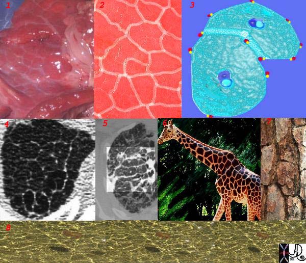
This is a series of images demonstrating the shape of the secondary lobule. The first image (1) is a post mortem specimen with congested lungs showing the interlobular septa, while the next (2), is an overlay of the septa in white showing their polygonal shape. The next drawing reveals side by side secondary lobules with central bronchovascular bundles and peripheral lymphovascular bundles. Image 4 is a CT image through the apex of the lung showing thickened secondary lobules in a patient with mild emphysema, and 5 shows marked thickening of the interlobular septa in a patient with end stage sarcoidosis. 6,7,8, show the shape of the secondary lobules in the skin of a giraffe, the bark of a pine, and the ripples of the water respectively. Courtesy Ashley Davidoff MD. 31866collage code lungs pulmonary secondary lobule interlobular septa thick thickened thickening giraffe drawing grosspathology CTscan lymphatics imaging CTscan collage drawing

This is a series of images demonstrating the shape of the secondary lobule. The first image (1) is a post mortem specimen with congested lungs showing the interlobular septa, while the next (2), is an overlay of the septa in white showing their polygonal shape. The next drawing reveals side by side secondary lobules with central bronchovascular bundles and peripheral lymphovascular bundles. Image 4 is a CT image through the apex of the lung showing thickened secondary lobules in a patient with mild emphysema, and 5 shows marked thickening of the interlobular septa in a patient with end stage sarcoidosis. 6,7,8, show the shape of the secondary lobules in the skin of a giraffe, the bark of a pine, and the ripples of the water respectively. Courtesy Ashley Davidoff MD. 31866collage code lungs pulmonary secondary lobule interlobular septa thick thickened thickening giraffe drawing grosspathology CTscan lymphatics imaging CTscan collage drawing