42651c

This collage reflects the range of the respiratory system from the macroscopic and anatomic to the microscopic - a continuum of structure. Image 2 is a post-mortem specimen taken from the front and slightly above. It shows the trachea and bronchi supplying the two lungs above, with the aortic arch and cardiac structures in the middle and below. Note how pink the lungs are in this specimen from an unfortunate baby with congenital heart disease. Image 3, the chest X-ray, shows the lucent lungs within the thoracic cavity while image 4 is a diagram of the trilobed right lung and the bilobed left lung. Two respiratory units of the lung are shown in the next image each called a pulmonary lobule (5). The lobule consists of a central bronchiole (light blue) and pulmonary arteriole (dark blue), surrounded by the air filled acinus (teal) with its peripheral venules. (red) The acinus is magnified in the next image (6), showing first the tubular terminal bronchiole branching into the respiratory bronchioles, alveolar sacs, and finally the grape like alveoli. The organization of the connective tissues of the lung is shown in image 7. Finally we get down to the grapes or alveoli of the lung with surrounding vessels (8), and a single alveolus is seen in 9. It seems a long way for the air to travel but the system can deliver the air to and from the outside in a single breath, and exchange the gases at the capillary level even more rapidly. It is a remarkable system. Courtesy Ashley Davidoff MD 42651c code lung chest Davidoff art

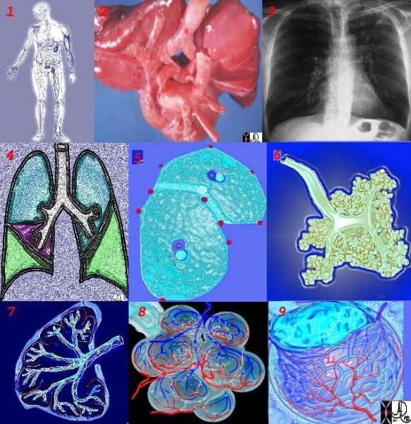
This collage reflects the range of the respiratory system from the macroscopic and anatomic to the microscopic – a continuum of structure. Image 2 is a post-mortem specimen taken from the front and slightly above. It shows the trachea and bronchi supplying the two lungs above, with the aortic arch and cardiac structures in the middle and below. Note how pink the lungs are in this specimen from an unfortunate baby with congenital heart disease. Image 3, the chest X-ray, shows the lucent lungs within the thoracic cavity while image 4 is a diagram of the trilobed right lung and the bilobed left lung. Two respiratory units of the lung are shown in the next image each called a pulmonary lobule (5). The lobule consists of a central bronchiole (light blue) and pulmonary arteriole (dark blue), surrounded by the air filled acinus (teal) with its peripheral venules. (red) The acinus is magnified in the next image (6), showing first the tubular terminal bronchiole branching into the respiratory bronchioles, alveolar sacs, and finally the grape like alveoli. The organization of the connective tissues of the lung is shown in image 7. Finally we get down to the grapes or alveoli of the lung with surrounding vessels (8), and a single alveolus is seen in 9. It seems a long way for the air to travel but the system can deliver the air to and from the outside in a single breath, and exchange the gases at the capillary level even more rapidly. It is a remarkable system. Courtesy Ashley Davidoff MD 42651c code lung chest Davidoff art