
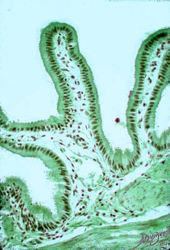
Normal Gallbladder Histology
This is a medium power photomicrograph showing how delicate the mucosal fronds are. Each frond consists of a single layer of columnar epithelium covering a core of capillaries and a few fibroblasts. The empty part of the picture would be the gallbladder lumen, and on the opposite side parts of two muscle bundles can be seen.